

 Français
 Español
 Products
 About Us


Mucus specimens are crucial in diagnosing various respiratory and gastrointestinal conditions. Collecting and analyzing these specimens properly can lead to early detection of infections, inflammation, and other abnormalities. This comprehensive guide explains the best practices for mucus specimen collection, laboratory analysis, and result interpretation.
| |
| |
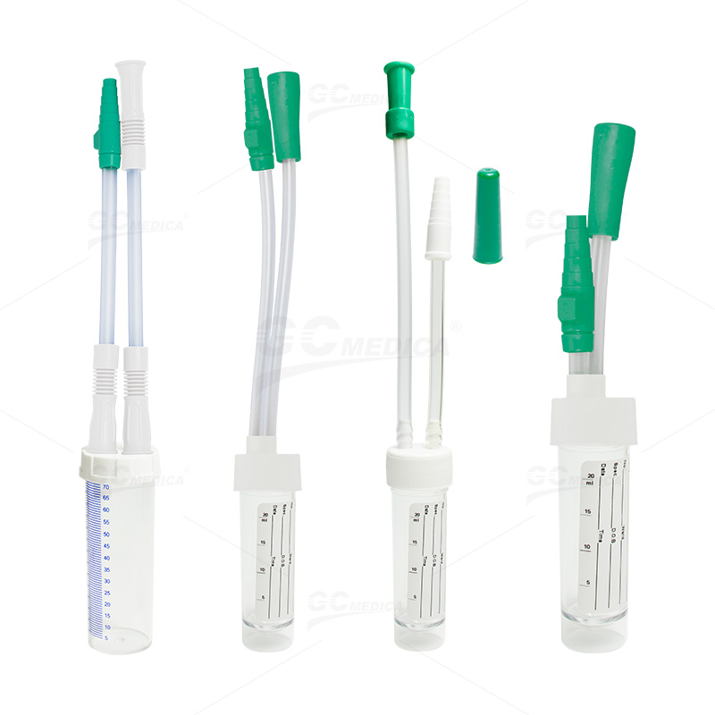
| Mucus Specimen Trap 25ml/70ml |


Collection of Mucus Specimens
The first step in obtaining an accurate diagnosis is proper collection. Depending on the suspected condition, mucus specimens can be collected from the respiratory tract or gastrointestinal system. Here are key collection guidelines:
Patient Preparation: Instruct patients to avoid eating, drinking, or smoking before sample collection if applicable. Clear communication helps in minimizing contamination.
Collection Techniques: For respiratory specimens, ask the patient to cough deeply into a sterile container. In some cases, induced sputum or bronchoalveolar lavage might be necessary. For gastrointestinal mucus, collection may involve endoscopic techniques.
Sterility: Use sterile containers and equipment to prevent cross-contamination. Label each specimen clearly with patient details and the time of collection.
Laboratory Analysis
Once collected, mucus specimens undergo a series of laboratory tests to identify pathogens, inflammatory markers, or other cellular abnormalities. Key steps include:
Microscopic Examination: Laboratory technicians use light microscopy to identify cellular components, bacteria, or fungal elements in the mucus.
Culture and Sensitivity: Culturing the specimen helps in isolating and identifying bacterial or viral pathogens. This step is critical for determining the appropriate antibiotic treatment.
Biochemical and Molecular Tests: Advanced techniques such as PCR (polymerase chain reaction) allow for the detection of specific genetic markers, providing a more accurate diagnosis. Enzyme-linked immunosorbent assays (ELISAs) can detect specific antigens or antibodies present in the sample.
Interpretation of Results
The interpretation of mucus specimen results is vital for guiding treatment decisions. Here are some factors to consider:
Normal vs. Abnormal Findings: A healthy mucus specimen typically contains minimal inflammatory cells and no significant pathogenic organisms. Elevated levels of white blood cells, presence of pathogens, or abnormal cell structures indicate possible infections or inflammatory conditions.
Clinical Correlation: Laboratory results must be interpreted in conjunction with the patient’s symptoms and medical history. For example, increased mucus viscosity and bacterial growth might correlate with chronic bronchitis, while viral infections could display different inflammatory patterns.
Follow-Up Testing: Sometimes, further tests such as imaging studies or repeat specimen collection might be necessary to confirm a diagnosis or monitor treatment efficacy.
Best Practices and Recommendations
Timely Processing: Mucus specimens should be processed as soon as possible after collection to maintain sample integrity. Delays can result in the degradation of cells and pathogens.
Quality Control: Regularly review collection and processing protocols to ensure consistency and accuracy in laboratory results.
Patient Education: Educate patients on the importance of following pre-collection instructions and informing healthcare providers about any factors that might affect the test results, such as recent medications or dietary habits.
Conclusion
Accurate mucus specimen collection, thorough laboratory analysis, and careful result interpretation are essential for effective diagnosis and treatment planning. By adhering to best practices, healthcare providers can improve diagnostic accuracy, enhance patient care, and ensure timely treatment interventions. This comprehensive approach not only aids in managing respiratory and gastrointestinal conditions but also contributes to better overall patient outcomes.


